CRISPR base editing gives measure of hope to people with muscular dystrophy

Next year, a neurologist will test CRISPR base editing in a trial of five people with muscular dystrophy to see if their muscles accept corrected cells and whether they multiply and take over the function of damaged cells.
When Martin Weber climbs the steps to his apartment on the fifth floor in Munich, an attentive observer might notice that he walks a little unevenly. “That’s because my calf muscles were the first to lose strength,” Weber explains.
About three years ago, the now 19-year-old university student realized that he suddenly had trouble keeping up with his track team at school. At tennis tournaments, he seemed to lose stamina after the first hour. “But it was still within the norm,” he says. “So it took a while before I noticed something was seriously wrong.” A blood test showed highly elevated liver markers. His parents feared he had liver cancer until a week-long hospital visit and scores of tests led to a diagnosis: hereditary limb-girdle muscular dystrophy, an incurable genetic illness that causes muscles to deteriorate.
As you read this text, you will surely use several muscles without being aware of them: Your heart muscle pumps blood through your arteries, your eye muscles let you follow the words in this sentence, and your hand muscles hold the tablet or cell phone. Muscles make up 40 percent of your body weight; we usually have 656 of them. Now imagine they are slowly losing their strength. No training, no protein shake can rebuild their function.
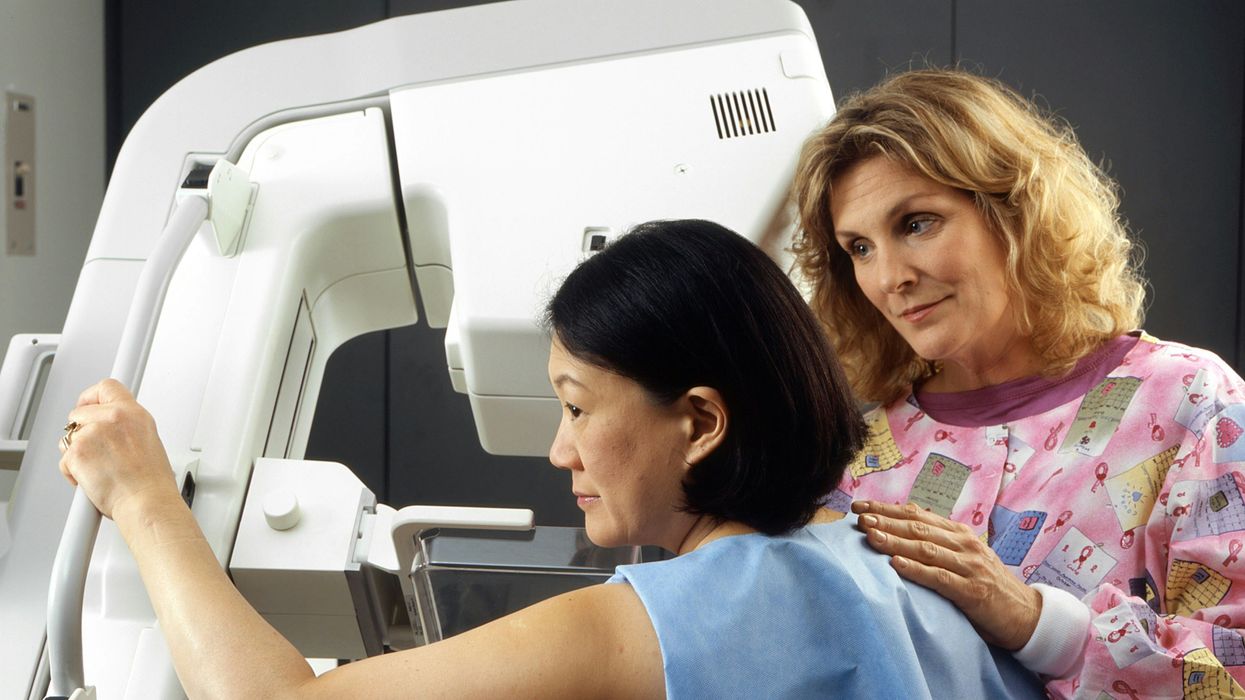
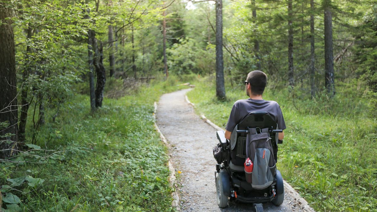
This is the reality for most people in Simone Spuler’s outpatient clinic at the Charité Hospital in Berlin, Germany: Almost all of her 2,500 patients have muscular dystrophy, a progressive illness striking mostly young people. Muscle decline leads to a wheelchair and, eventually, an early death due to a heart attack or the inability to breathe. In Germany alone, 300,000 people live with this illness, the youngest barely a year old. The CDC estimates that its most common form, Duchenne, affects 1 in every 3,500 to 6,000 male births each year in the United States.
The devastating progression of the disease is what motivates Spuler and her team of 25 scientists to find a cure. In 2019, they made a spectacular breakthrough: For the first time, they successfully used mRNA to introduce the CRISPR-Cas9 tool into human muscle stem cells to repair the dystrophy. “It’s really just one tiny molecule that doesn’t work properly,” Spuler explains.
CRISPR-Cas9 is a technology that lets scientists select and alter parts of the genome. It’s still comparatively new but has advanced quickly since its discovery in the early 2010s. “We now have the possibility to repair certain mutations with genetic editing,” Spuler says. “It’s pure magic.”
She projects a warm, motherly air and a professional calm that inspires trust from her patients. She needs these qualities because the 60-year-old neurologist has one of the toughest jobs in the world: All day long, patients with the incurable diagnosis of muscular dystrophy come to her clinic, and she watches them decline over the years. “Apart from physiotherapy, there is nothing we can recommend right now,” she says. That motivated her early in her career, when she met her first patients at the Max Planck Institute for Neurobiology near Munich in the 1990s. “I knew I had 30, 40 years to find something.”
She learned from the luminaries of her profession with postdocs at the University of California San Diego, Harvard and Johns Hopkins, before serving as a clinical fellow at the Mayo Clinic. In 2005, the Charité offered her the opportunity to establish a specialized clinic for myasthenia, or muscular weakness. An important influence on Spuler, she says, has been the French microbiologist Emmanuelle Charpentier, who received the Nobel Prize in 2020 along with Jennifer Doudna for their CRISPR research, and has worked in Berlin since 2015.
When CRISPR was first introduced, it was mainly used to cut through DNA. However, the cut can lead to undesired side effects. For the muscle stem cells, Spuler now uses a base editor to repair the damaged molecule with super fine scissors or tweezers.
“Apart from physiotherapy, there is nothing we can recommend right now,” Spuler says about her patients with limb-girdle muscular dystrophy.
Pablo Castagnola
Last year, she proved that the method works in mice. Injecting repaired cells into the rodents led to new muscle fibers and, in 2021 and 2022, she passed the first safety meetings with the Paul-Ehrlich Institute, which is responsible for approving human gene editing trials in Germany. She raised the nearly four million Euros needed to test the new method in the first clinical trial in humans with limb-girdle muscular dystrophy, beginning with one muscle that can easily be measured, such as the biceps.
This spring, Weber and his parents drove the 400 miles from Munich to Berlin. At Spuler’s lab, her team took a biopsy from muscles in his left arm. The first two steps – extraction and repair in a culture dish – went according to plan; Spuler was able to repair the mutation in Weber’s cells outside his body.
Next year, Weber will be the youngest participant when Spuler starts to test the method in a trial of five people “in vivo,” inside their bodies. This will be the real moment of truth: Will the participants’ muscles accept the corrected cells? Will the cells multiply and take over the function of damaged cells, just like Spuler was able to do in her lab with the rodents?
The effort is costly and complex. “The biggest challenge is to make absolutely sure that we don’t harm the patient,” Spuler says. This means scanning their entire genomes, “so we don’t accidentally damage or knock out an important gene.”
Weber, who asked not to be identified by his real name, is looking forward to the trial and he feels confident that “the risks are comparatively small because the method will only be applied to one muscle. The worst that can happen is that it doesn’t work. But in the best case, the muscle function will improve.”
He was so impressed with the Charité scientists that he decided to study biology at his university. He’s read extensively about CRISPR, so he understands why he has three healthy siblings. “That’s the statistics,” the biologist in training explains. “You get two sets of genes from each parent, and you have to get two faulty mutations to have muscular dystrophy. So we fit the statistics exactly: One of us four kids inherited the mutation.”
It was his mother, a college teacher, and father, a physicist by training, who heard about Spuler’s research. Even though Weber does not live at home anymore, having a chronically ill son is nearly a full-time job for his mother, Annette. The Berlin visit and the trial are financed separately through private sponsors, but the fights with Weber’s health insurance are frustrating and time-consuming. “Physiotherapy is the only thing that helps a bit,” Weber says, “and yet, they fought us on approving it every step of the way.”
Spuler does not want to evoke unrealistic expectations. “Patients who are wheelchair-bound won’t suddenly get up and walk."
Her son continues to exercise as much as possible. Riding his bicycle to the university has become too difficult, so he got an e-scooter. He had to give up competitive tennis because he does not have the stamina for a two-hour match, but he can still play with his dad or his buddies for an hour. His closest friends know about the diagnosis. “They help me, for instance, to lift something heavy because I can’t do that anymore,” Weber says.
The family was elated to find medical support at the Munich Muscle Center by the German Alliance for Muscular Patients and then at Spuler’s clinic in Berlin. “When you hear that this is a progressive illness with no chance of improvement, your world collapses as a parent,” Annette Weber says. “And then all of a sudden, there is this woman who sees scientific progress as an opportunity. Even just to be able to participate in the study is fantastic.”
Spuler does not want to evoke unrealistic expectations. “Patients who are wheelchair-bound won’t suddenly get up and walk,” she says. After all, she will start by applying the gene editor to only one muscle, “but it would be a big step if even a small muscle that is essential to grip something, or to swallow, regains function.”
Weber agrees. “I understand that I won’t regain 100 percent of my muscle function but even a small improvement or at least halting the deterioration is the goal.”
And yet, Spuler and others are ultimately searching for a true solution. In a separate effort, Massachusetts-based biotech company Sarepta announced this month it will seek expedited regulators’ approval to treat Duchenne patients with its investigational gene therapy. Unlike Spuler’s methods, Sarepta focuses specifically on the Duchenne form of muscular dystrophy, and it uses an adeno-assisted virus to deliver the therapy.
Spuler’s vision is to eventually apply gene editing to the entire body of her patients. To speed up the research, she and a colleague founded a private research company, Myopax. If she is able to prove that the body accepts the edited cells, the technique could be used for other monogenetic illnesses as well. “When we speak of genetic editing, many are scared and say, oh no, this is God’s work,” says Spuler. But she sees herself as a mechanic, not a divine being. “We really just exchange a molecule, that’s it.”
If everything goes well, Weber hopes that ten years from now, he will be the one taking biopsies from the next generation of patients and repairing their genes.
Tiny, tough “water bears” may help bring new vaccines and medicines to sub-Saharan Africa
Tardigrades can completely dehydrate and later rehydrate themselves, a survival trick that scientists are harnessing to preserve medicines in hot temperatures.
Microscopic tardigrades, widely considered to be some of the toughest animals on earth, can survive for decades without oxygen or water and are thought to have lived through a crash-landing on the moon. Also known as water bears, they survive by fully dehydrating and later rehydrating themselves – a feat only a few animals can accomplish. Now scientists are harnessing tardigrades’ talents to make medicines that can be dried and stored at ambient temperatures and later rehydrated for use—instead of being kept refrigerated or frozen.
Many biologics—pharmaceutical products made by using living cells or synthesized from biological sources—require refrigeration, which isn’t always available in many remote locales or places with unreliable electricity. These products include mRNA and other vaccines, monoclonal antibodies and immuno-therapies for cancer, rheumatoid arthritis and other conditions. Cooling is also needed for medicines for blood clotting disorders like hemophilia and for trauma patients.
Formulating biologics to withstand drying and hot temperatures has been the holy grail for pharmaceutical researchers for decades. It’s a hard feat to manage. “Biologic pharmaceuticals are highly efficacious, but many are inherently unstable,” says Thomas Boothby, assistant professor of molecular biology at University of Wyoming. Therefore, during storage and shipping, they must be refrigerated at 2 to 8 degrees Celsius (35 to 46 degrees Fahrenheit). Some must be frozen, typically at -20 degrees Celsius, but sometimes as low -90 degrees Celsius as was the case with the Pfizer Covid vaccine.
For Covid, fewer than 73 percent of the global population received even one dose. The need for refrigerated or frozen handling was partially to blame.
The costly cold chain
The logistics network that ensures those temperature requirements are met from production to administration is called the cold chain. This cold chain network is often unreliable or entirely lacking in remote, rural areas in developing nations that have malfunctioning electrical grids. “Almost all routine vaccines require a cold chain,” says Christopher Fox, senior vice president of formulations at the Access to Advanced Health Institute. But when the power goes out, so does refrigeration, putting refrigerated or frozen medical products at risk. Consequently, the mRNA vaccines developed for Covid-19 and other conditions, as well as more traditional vaccines for cholera, tetanus and other diseases, often can’t be delivered to the most remote parts of the world.
To understand the scope of the challenge, consider this: In the U.S., more than 984 million doses of Covid-19 vaccine have been distributed so far. Each one needed refrigeration that, even in the U.S., proved challenging. Now extrapolate to all vaccines and the entire world. For Covid, fewer than 73 percent of the global population received even one dose. The need for refrigerated or frozen handling was partially to blame.
Globally, the cold chain packaging market is valued at over $15 billion and is expected to exceed $60 billion by 2033.
Adobe Stock
Freeze-drying, also called lyophilization, which is common for many vaccines, isn’t always an option. Many freeze-dried vaccines still need refrigeration, and even medicines approved for storage at ambient temperatures break down in the heat of sub-Saharan Africa. “Even in a freeze-dried state, biologics often will undergo partial rehydration and dehydration, which can be extremely damaging,” Boothby explains.
The cold chain is also very expensive to maintain. The global pharmaceutical cold chain packaging market is valued at more than $15 billion, and is expected to exceed $60 billion by 2033, according to a report by Future Market Insights. This cost is only expected to grow. According to the consulting company Accenture, the number of medicines that require the cold chain are expected to grow by 48 percent, compared to only 21 percent for non-cold-chain therapies.
Tardigrades to the rescue
Tardigrades are only about a millimeter long – with four legs and claws, and they lumber around like bears, thus their nickname – but could provide a big solution. “Tardigrades are unique in the animal kingdom, in that they’re able to survive a vast array of environmental insults,” says Boothby, the Wyoming professor. “They can be dried out, frozen, heated past the boiling point of water and irradiated at levels that are thousands of times more than you or I could survive.” So, his team is gradually unlocking tardigrades’ survival secrets and applying them to biologic pharmaceuticals to make them withstand both extreme heat and desiccation without losing efficacy.
Boothby’s team is focusing on blood clotting factor VIII, which, as the name implies, causes blood to clot. Currently, Boothby is concentrating on the so-called cytoplasmic abundant heat soluble (CAHS) protein family, which is found only in tardigrades, protecting them when they dry out. “We showed we can desiccate a biologic (blood clotting factor VIII, a key clotting component) in the presence of tardigrade proteins,” he says—without losing any of its effectiveness.
The researchers mixed the tardigrade protein with the blood clotting factor and then dried and rehydrated that substance six times without damaging the latter. This suggests that biologics protected with tardigrade proteins can withstand real-world fluctuations in humidity.
Furthermore, Boothby’s team found that when the blood clotting factor was dried and stabilized with tardigrade proteins, it retained its efficacy at temperatures as high as 95 degrees Celsius. That’s over 200 degrees Fahrenheit, much hotter than the 58 degrees Celsius that the World Meteorological Organization lists as the hottest recorded air temperature on earth. In contrast, without the protein, the blood clotting factor degraded significantly. The team published their findings in the journal Nature in March.
Although tardigrades rarely live more than 2.5 years, they have survived in a desiccated state for up to two decades, according to Animal Diversity Web. This suggests that tardigrades’ CAHS protein can protect biologic pharmaceuticals nearly indefinitely without refrigeration or freezing, which makes it significantly easier to deliver them in locations where refrigeration is unreliable or doesn’t exist.
The tricks of the tardigrades
Besides the CAHS proteins, tardigrades rely on a type of sugar called trehalose and some other protectants. So, rather than drying up, their cells solidify into rigid, glass-like structures. As that happens, viscosity between cells increases, thereby slowing their biological functions so much that they all but stop.
Now Boothby is combining CAHS D, one of the proteins in the CAHS family, with trehalose. He found that CAHS D and trehalose each protected proteins through repeated drying and rehydrating cycles. They also work synergistically, which means that together they might stabilize biologics under a variety of dry storage conditions.
“We’re finding the protective effect is not just additive but actually is synergistic,” he says. “We’re keen to see if something like that also holds true with different protein combinations.” If so, combinations could possibly protect against a variety of conditions.
Commercialization outlook
Before any stabilization technology for biologics can be commercialized, it first must be approved by the appropriate regulators. In the U.S., that’s the U.S. Food and Drug Administration. Developing a new formulation would require clinical testing and vast numbers of participants. So existing vaccines and biologics likely won’t be re-formulated for dry storage. “Many were developed decades ago,” says Fox. “They‘re not going to be reformulated into thermo-stable vaccines overnight,” if ever, he predicts.
Extending stability outside the cold chain, even for a few days, can have profound health, environmental and economic benefits.
Instead, this technology is most likely to be used for the new products and formulations that are just being created. New and improved vaccines will be the first to benefit. Good candidates include the plethora of mRNA vaccines, as well as biologic pharmaceuticals for neglected diseases that affect parts of the world where reliable cold chain is difficult to maintain, Boothby says. Some examples include new, more effective vaccines for malaria and for pathogenic Escherichia coli, which causes diarrhea.
Tallying up the benefits
Extending stability outside the cold chain, even for a few days, can have profound health, environmental and economic benefits. For instance, MenAfriVac, a meningitis vaccine (without tardigrade proteins) developed for sub-Saharan Africa, can be stored at up to 40 degrees Celsius for four days before administration. “If you have a few days where you don’t need to maintain the cold chain, it’s easier to transport vaccines to remote areas,” Fox says, where refrigeration does not exist or is not reliable.
Better health is an obvious benefit. MenAfriVac reduced suspected meningitis cases by 57 percent in the overall population and more than 99 percent among vaccinated individuals.
Lower healthcare costs are another benefit. One study done in Togo found that the cold chain-related costs increased the per dose vaccine price up to 11-fold. The ability to ship the vaccines using the usual cold chain, but transporting them at ambient temperatures for the final few days cut the cost in half.
There are environmental benefits, too, such as reducing fuel consumption and greenhouse gas emissions. Cold chain transports consume 20 percent more fuel than non-cold chain shipping, due to refrigeration equipment, according to the International Trade Administration.
A study by researchers at Johns Hopkins University compared the greenhouse gas emissions of the new, oral Vaxart COVID-19 vaccine (which doesn’t require refrigeration) with four intramuscular vaccines (which require refrigeration or freezing). While the Vaxart vaccine is still in clinical trials, the study found that “up to 82.25 million kilograms of CO2 could be averted by using oral vaccines in the U.S. alone.” That is akin to taking 17,700 vehicles out of service for one year.
Although tardigrades’ protective proteins won’t be a component of biologic pharmaceutics for several years, scientists are proving that this approach is viable. They are hopeful that a day will come when vaccines and biologics can be delivered anywhere in the world without needing refrigerators or freezers en route.
Jamie Rettinger with his now fiance Amie Purnel-Davis, who helped him through the clinical trial.
Jamie Rettinger was still in his thirties when he first noticed a tiny streak of brown running through the thumbnail of his right hand. It slowly grew wider and the skin underneath began to deteriorate before he went to a local dermatologist in 2013. The doctor thought it was a wart and tried scooping it out, treating the affected area for three years before finally removing the nail bed and sending it off to a pathology lab for analysis.
"I have some bad news for you; what we removed was a five-millimeter melanoma, a cancerous tumor that often spreads," Jamie recalls being told on his return visit. "I'd never heard of cancer coming through a thumbnail," he says. None of his doctors had ever mentioned it either. "I just thought I was being treated for a wart." But nothing was healing and it continued to bleed.
A few months later a surgeon amputated the top half of his thumb. Lymph node biopsy tested negative for spread of the cancer and when the bandages finally came off, Jamie thought his medical issues were resolved.
Melanoma is the deadliest form of skin cancer. About 85,000 people are diagnosed with it each year in the U.S. and more than 8,000 die of the cancer when it spreads to other parts of the body, according to the Centers for Disease Control and Prevention (CDC).
There are two peaks in diagnosis of melanoma; one is in younger women ages 30-40 and often is tied to past use of tanning beds; the second is older men 60+ and is related to outdoor activity from farming to sports. Light-skinned people have a twenty-times greater risk of melanoma than do people with dark skin.
"When I graduated from medical school, in 2005, melanoma was a death sentence" --Diwakar Davar.
Jamie had a follow up PET scan about six months after his surgery. A suspicious spot on his lung led to a biopsy that came back positive for melanoma. The cancer had spread. Treatment with a monoclonal antibody (nivolumab/Opdivo®) didn't prove effective and he was referred to the UPMC Hillman Cancer Center in Pittsburgh, a four-hour drive from his home in western Ohio.
An alternative monoclonal antibody treatment brought on such bad side effects, diarrhea as often as 15 times a day, that it took more than a week of hospitalization to stabilize his condition. The only options left were experimental approaches in clinical trials.
Early research
"When I graduated from medical school, in 2005, melanoma was a death sentence" with a cure rate in the single digits, says Diwakar Davar, 39, an oncologist at UPMC Hillman Cancer Center who specializes in skin cancer. That began to change in 2010 with introduction of the first immunotherapies, monoclonal antibodies, to treat cancer. The antibodies attach to PD-1, a receptor on the surface of T cells of the immune system and on cancer cells. Antibody treatment boosted the melanoma cure rate to about 30 percent. The search was on to understand why some people responded to these drugs and others did not.
At the same time, there was a growing understanding of the role that bacteria in the gut, the gut microbiome, plays in helping to train and maintain the function of the body's various immune cells. Perhaps the bacteria also plays a role in shaping the immune response to cancer therapy.
One clue came from genetically identical mice. Animals ordered from different suppliers sometimes responded differently to the experiments being performed. That difference was traced to different compositions of their gut microbiome; transferring the microbiome from one animal to another in a process known as fecal transplant (FMT) could change their responses to disease or treatment.
When researchers looked at humans, they found that the patients who responded well to immunotherapies had a gut microbiome that looked like healthy normal folks, but patients who didn't respond had missing or reduced strains of bacteria.
Davar and his team knew that FMT had a very successful cure rate in treating the gut dysbiosis of Clostridioides difficile, a persistant intestinal infection, and they wondered if a fecal transplant from a patient who had responded well to cancer immunotherapy treatment might improve the cure rate of patients who did not originally respond to immunotherapies for melanoma.
The ABCDE of melanoma detection
Adobe Stock
Clinical trial
"It was pretty weird, I was totally blasted away. Who had thought of this?" Jamie first thought when the hypothesis was explained to him. But Davar's explanation that the procedure might restore some of the beneficial bacterial his gut was lacking, convinced him to try. He quickly signed on in October 2018 to be the first person in the clinical trial.
Fecal donations go through the same safety procedures of screening for and inactivating diseases that are used in processing blood donations to make them safe for transfusion. The procedure itself uses a standard hollow colonoscope designed to screen for colon cancer and remove polyps. The transplant is inserted through the center of the flexible tube.
Most patients are sedated for procedures that use a colonoscope but Jamie doesn't respond to those drugs: "You can't knock me out. I was watching them on the TV going up my own butt. It was kind of unreal at that point," he says. "There were about twelve people in there watching because no one had seen this done before."
A test two weeks after the procedure showed that the FMT had engrafted and the once-missing bacteria were thriving in his gut. More importantly, his body was responding to another monoclonal antibody (pembrolizumab/Keytruda®) and signs of melanoma began to shrink. Every three months he made the four-hour drive from home to Pittsburgh for six rounds of treatment with the antibody drug.
"We were very, very lucky that the first patient had a great response," says Davar. "It allowed us to believe that even though we failed with the next six, we were on the right track. We just needed to tweak the [fecal] cocktail a little better" and enroll patients in the study who had less aggressive tumor growth and were likely to live long enough to complete the extensive rounds of therapy. Six of 15 patients responded positively in the pilot clinical trial that was published in the journal Science.
Davar believes they are beginning to understand the biological mechanisms of why some patients initially do not respond to immunotherapy but later can with a FMT. It is tied to the background level of inflammation produced by the interaction between the microbiome and the immune system. That paper is not yet published.
Surviving cancer
It has been almost a year since the last in his series of cancer treatments and Jamie has no measurable disease. He is cautiously optimistic that his cancer is not simply in remission but is gone for good. "I'm still scared every time I get my scans, because you don't know whether it is going to come back or not. And to realize that it is something that is totally out of my control."
"It was hard for me to regain trust" after being misdiagnosed and mistreated by several doctors he says. But his experience at Hillman helped to restore that trust "because they were interested in me, not just fixing the problem."
He is grateful for the support provided by family and friends over the last eight years. After a pause and a sigh, the ruggedly built 47-year-old says, "If everyone else was dead in my family, I probably wouldn't have been able to do it."
"I never hesitated to ask a question and I never hesitated to get a second opinion." But Jamie acknowledges the experience has made him more aware of the need for regular preventive medical care and a primary care physician. That person might have caught his melanoma at an earlier stage when it was easier to treat.
Davar continues to work on clinical studies to optimize this treatment approach. Perhaps down the road, screening the microbiome will be standard for melanoma and other cancers prior to using immunotherapies, and the FMT will be as simple as swallowing a handful of freeze-dried capsules off the shelf rather than through a colonoscopy. Earlier this year, the Food and Drug Administration approved the first oral fecal microbiota product for C. difficile, hopefully paving the way for more.
An older version of this hit article was first published on May 18, 2021