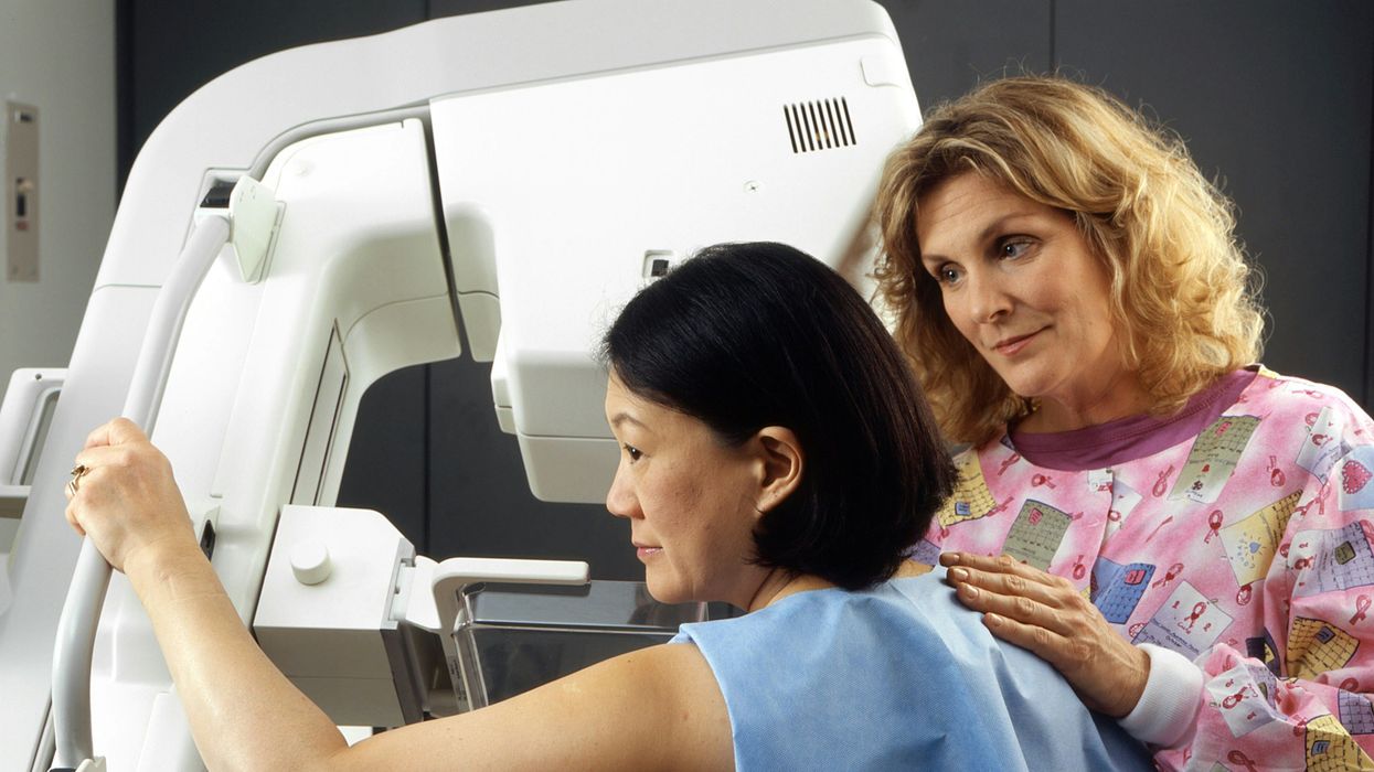
A woman checking her breast for the presence of concerning lumps.
Nancy Cappello was proactive. When she turned 36, she had a baseline mammogram, a standard medical recommendation in the late 1980s and early 1990s as a comparison tool for future screenings. At 40, Cappello started getting them annually.
Her breast surgeon estimated the cancer had been festering for four to five years under the radar of her annual mammograms.
Six weeks after her 11th-consecutive normal mammogram, she was diagnosed with Stage IIIc breast cancer.
A doctor felt a lump while doing a breast exam during her annual physical and a subsequent ultrasound detected cancer that had spread to 13 lymph nodes. That's when Cappello, then 51, learned she had dense breast tissue, making mammography less likely to detect tumors in her breasts.
She also discovered through her own research that she was among the 40 to 50 percent of women with dense breast tissue — almost half the female population — but medical protocol did not require physicians to inform women of their dense tissue status. If she had known, she said, she would have gotten an ultrasound every year in addition to a mammogram that could have detected the cancer much earlier. Cappello said her breast surgeon estimated the cancer had been festering for four to five years under the radar of her annual mammograms.
Although ultrasound as a cancer screening tool has been available for decades, technological advances are helping doctors find more invasive cancers in women with dense breasts, in turn giving women who know their tissue status the opportunity for earlier detection and treatment.
"We know that the gold standard for breast cancer screening is mammography, but in women with dense breast tissue, up to one third of breast cancers can be missed with this modality alone."
Dr. Georgia GiakoumisSpear, chief of the department of breast imaging at NorthShore University HealthSystem in suburban Chicago and assistant professor of radiology at the University of Chicago, has been a leader in developing standards for the use of new ultrasound technology. She is leading a study to develop more specific national guidelines around the use of Automated Whole Breast Tissue Ultrasound (ABUS), a non-invasive procedure in which sound waves are used to scan breast tissue while a patient lies on her back with her arm over her head.
Approved by the Food and Drug Administration in 2012, ABUS provides higher quality 3D images and faster delivery to provide more accurate results than past ultrasound technology. The scan does not involve radiation, and a practitioner can complete the process in 15 to 20 minutes, from patient preparation to image creation. NorthShore has been using ABUS since 2015, Dr. Spear said, and the technology can improve breast cancer detection in women with dense breasts by up to 55 percent.
"We know that the gold standard for breast cancer screening is mammography, but in women with dense breast tissue, up to one third of breast cancers can be missed with this modality alone," Spear says. "And when we supplement screening with ultrasound in this population of women, we have found a large number of cancers by ultrasound that are not visible on the mammogram."
Mammography should still be used as the first step for breast cancer detection, but if an initial mammogram shows that a patient has dense breast tissue, studies encourage discussion of additional screening with ultrasound.
On a mammogram, dense tissue appears white. So do cancerous masses, making them easy to miss.
A radiologist determines tissue density, according to the American College of Radiology's Breast Imaging Reporting and Data System (BI-RADS). "A" and "B" breast density categories designate ratios of mostly fatty, or non-dense tissues, while the "C" and "D" categories designate heterogeneously dense and extremely dense tissue, respectively. Such patients would be classified as having dense tissue. Younger women, women with lower levels of body fat and women undergoing hormone therapy are more likely to have C and D breast density.
On a mammogram, dense tissue appears white. So do cancerous masses, making them easy to miss. Fatty tissue, in comparison, appears black, making tumors easier to spot.
The FDA stated among its policy goals for 2018 that it's placing an improved focus on recognizing technological advances to help "ensure women get the most relevant, up-to-date information about their breast density, which is now recognized as a risk factor for breast cancer." An article in the March 2018 Journal of the American College of Radiology recommended supplemental screening for women with higher-than-average breast cancer risk, placing women with dense breast tissue in that category.
To be sure, some in the medical community are reluctant to push for ultrasounds, saying that a mammogram might be enough even if the woman has dense breast tissue. A patient is advised to discuss the option of ultrasound with her physician and they can decide from there.
Access to such information became political for Cappello after her diagnosis in 2004. She said that as she underwent six surgeries, a mastectomy, chemotherapy, radiation and hormone therapy, she asked doctors why they weren't required to inform women of their dense breast tissue status. Her dissatisfaction with their responses led to the formation of Are You Dense, Inc., an advocacy group aimed to inform women of their medical options while working to pass legislation mandating that women know their tissue status. Other legislation has focused on mandating insurance coverage for breast ultrasounds.

Nancy Cappello.
(Courtesy)
Cappello's work led Connecticut to become the first state to pass an information law in 2009, and 35 states now have similar requirements. Depending on the state, the law could mandate that certain language or information about breast density be included in the patient's mammogram results, or require physicians to tell women about dense tissue if their breast density falls in the BI-RADS categories C and D. Other states might require that patients be given general information about breast density and advice to discuss their options with a physician. (Note: There is a chart on Cappello's website that shows what laws exist – or don't – in each state.)
Through her site and social media, she's connected with other women who've lobbied for laws in their states, including Dr. Spear, who recently testified before legislative committees in Illinois as they considered companion bills. The Illinois legislation is expected to be signed into law this summer.
"There should be no excuses," Cappello says. "Women should have this information. There should be no concealing or hiding of her status."